Histopathology and Clinical Pathology exam preparation
Points
Necropsy of a rat
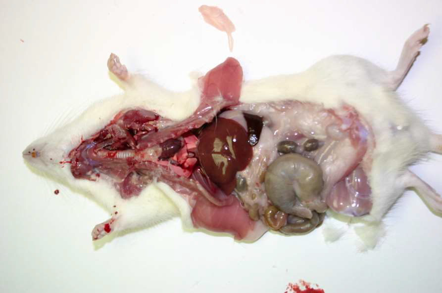
From slides


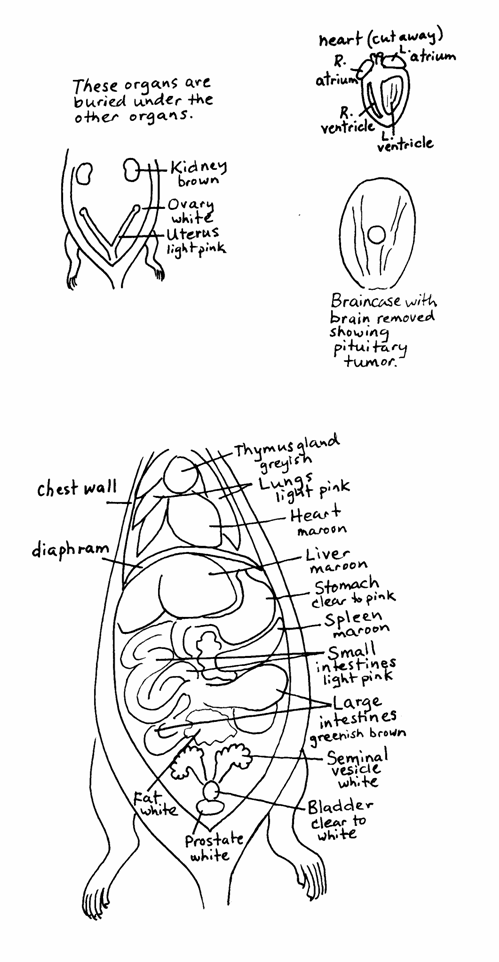
From http://ratfanclub.org/autopsy.html
HOW TO PREPARE THE SLIDES
Fixation
- Prevent autolysis
- Protect against microorganisms
- Increase the stability of the cells
Trimming
- Cut pieces of tissue into cassette
Dehydration, clearing & infiltration
Dehydration
Water in tissue removed
Gradually incresased concentration of ethanol
Clearing
- Ethanol is then removed
- Replaced by hydrophobic clearing agent (eg xylene)
Infiltration
- Xylene is then removed
- Replaced by paraffin wax
Embedding
- Infiltrated pieces of tissue embedded into blocks of paraffin wax
Sectioning
- Microtome is used for sectioning
- Commonly 4-6 micrometer thick sections
Mounting
- Sections collected in water bath (temperature)
- Transferred onto glass slides
Staining
Heamatoxylin and eosin (H&E)- Haematoxylin (basic dye) gives a dark blue stain of cell nuclei
- Eosin (acidic dye) stains the cytoplasm pinkish
Cover slip
Four basic types of tissues
- Epithelial
- Connective
- Muscle
- Nervous
Types of epithelium
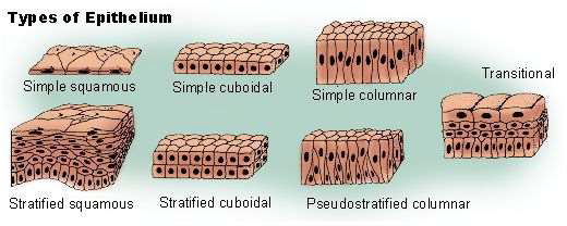
Types of connective tissue
- Fibrous (tendon)
- Skeletal (bone, cartilage)
- Fluid (blood, lymph)
Types of muscle
- Skeletal muscle
- Smooth muscle (In vessels, gastrointestinal, respiratory, urinary bladder, uterus)
- Cardiac muscle
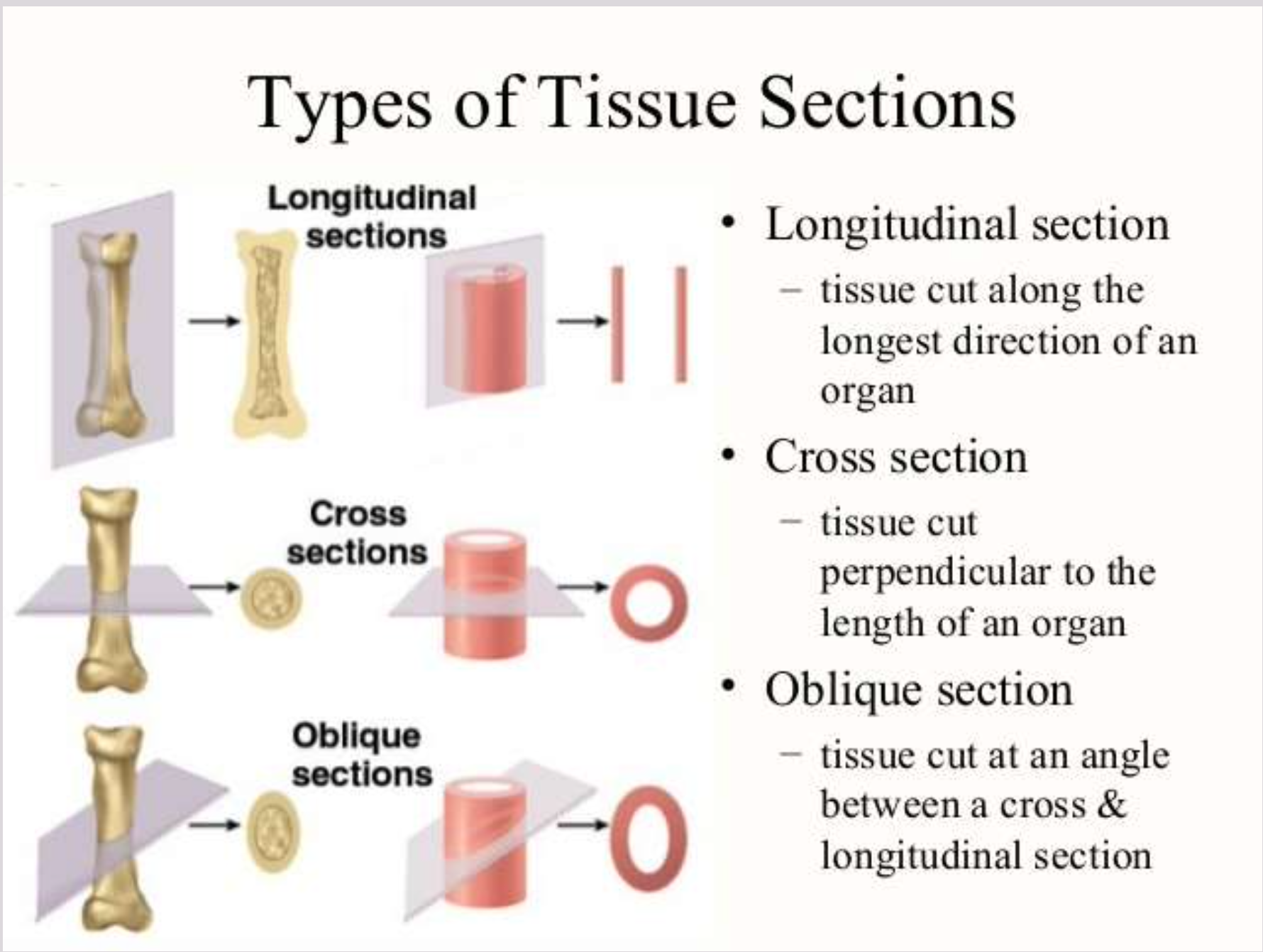
Types of tissue sections
Primary Organs
Liver
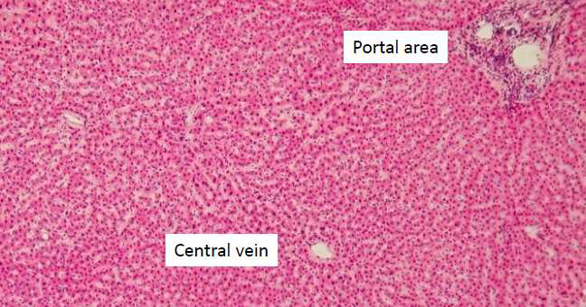
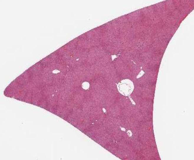
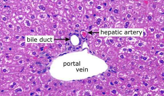
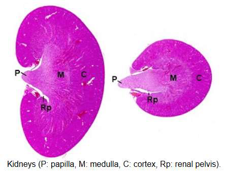
Kidney
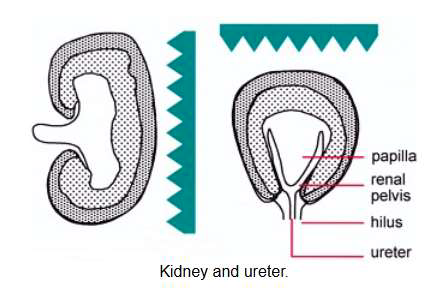
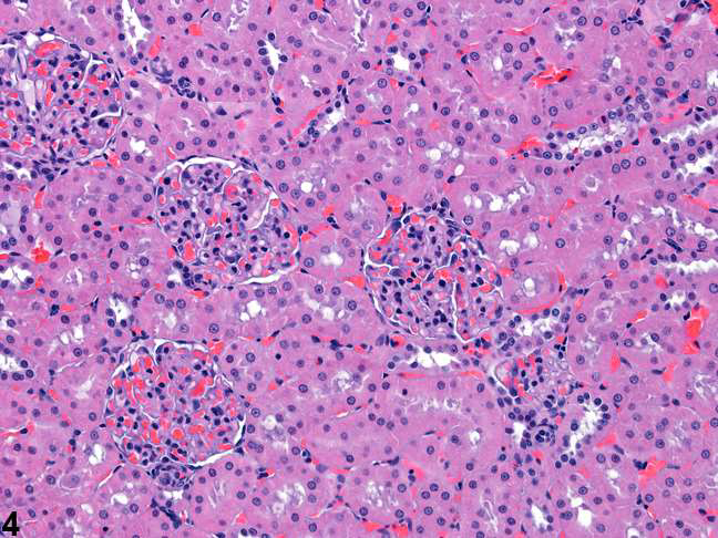
Renal tubules and glomeruli
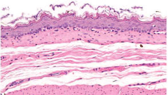
Stomach
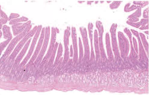
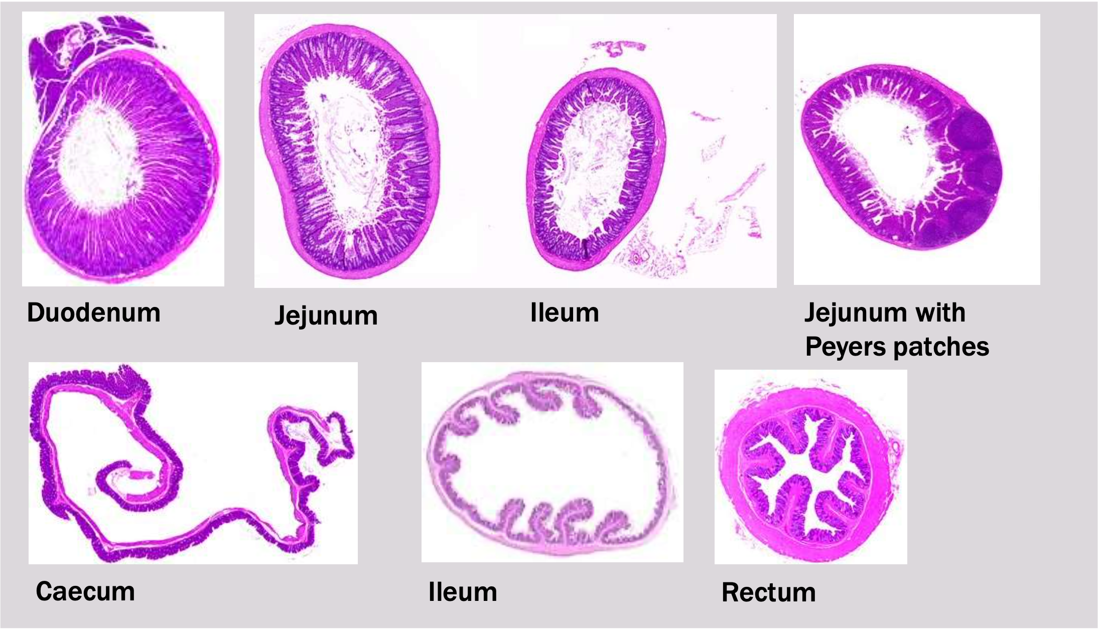
Intestines
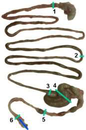
Small intestine
- duodenum
- jejunum
- ileum
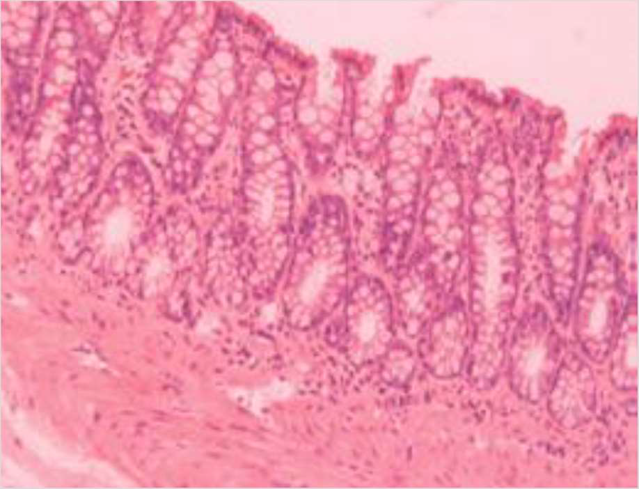
Large intestine
- cecum
- colon
- rectum
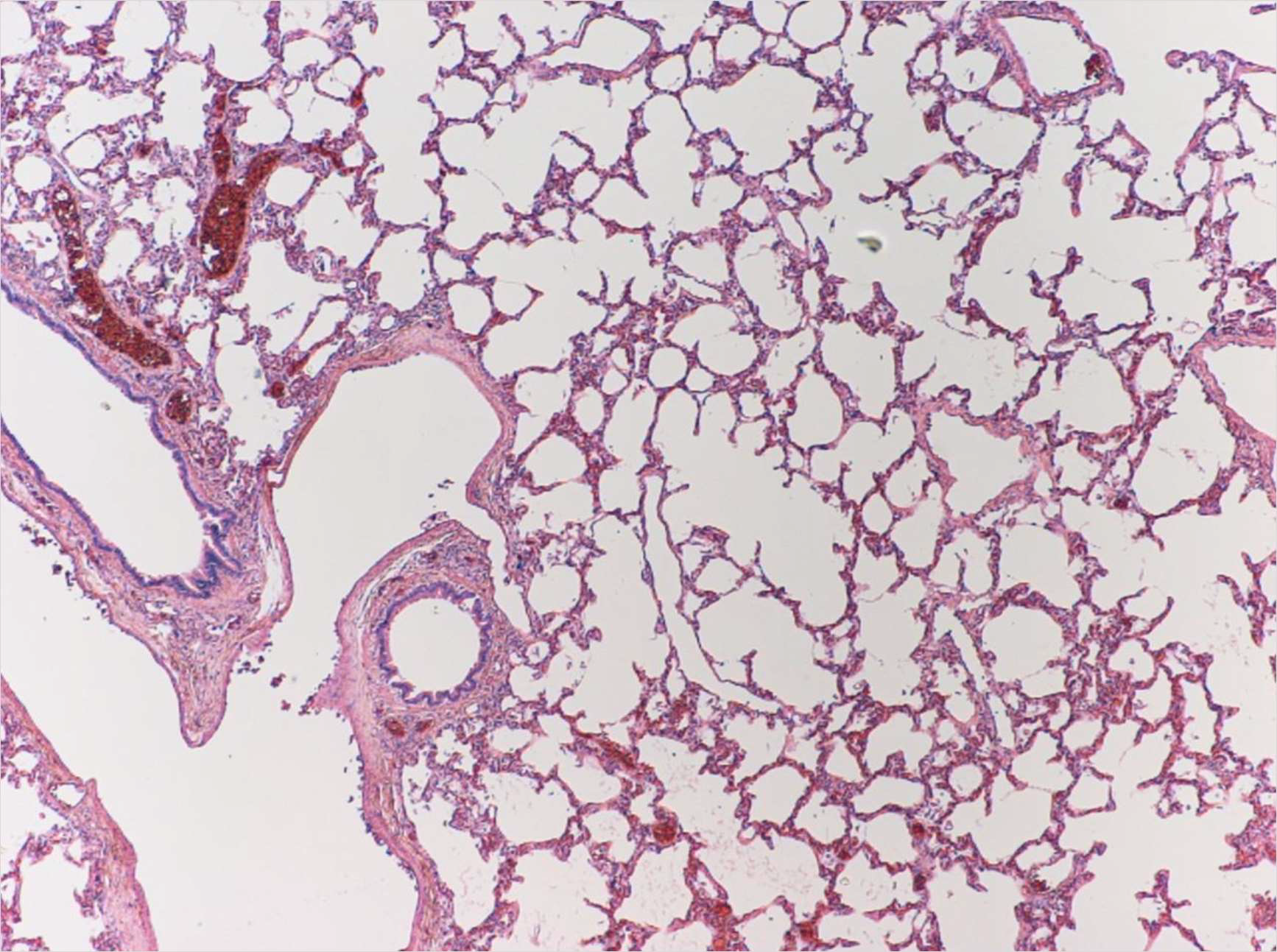
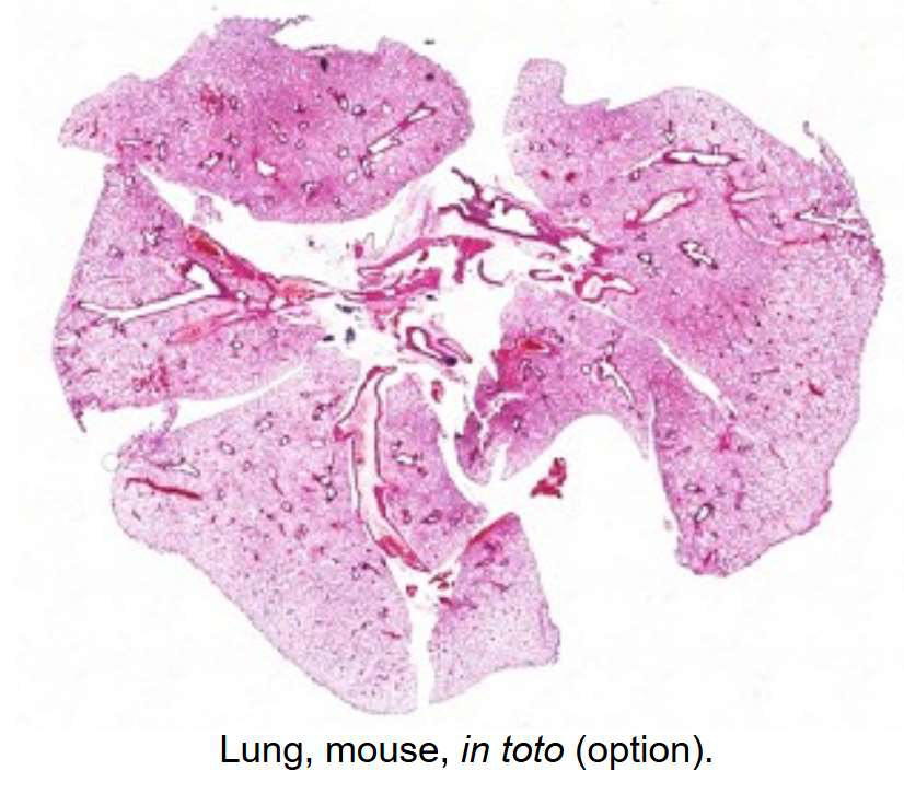
Lung
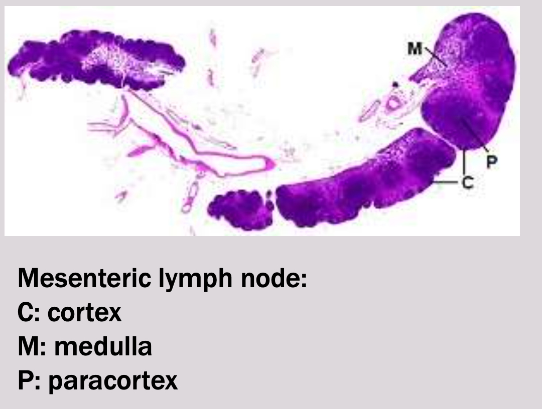
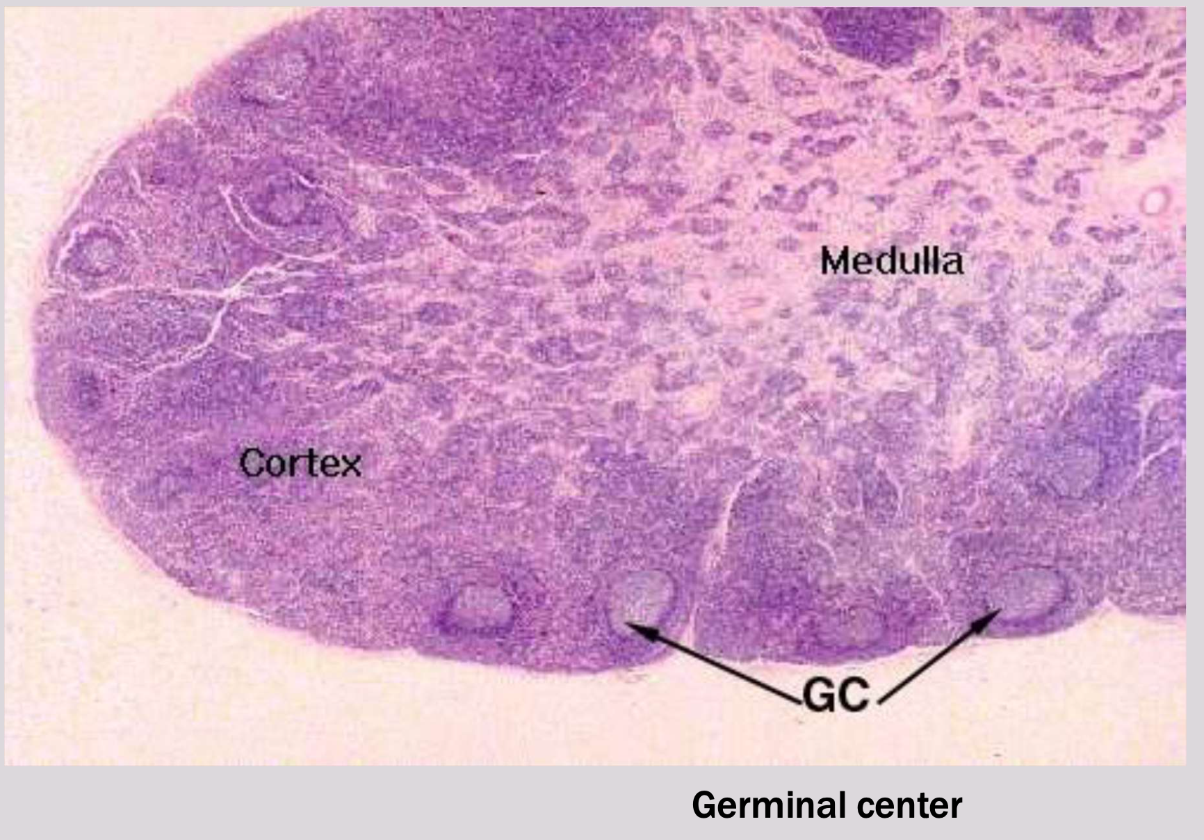
Lymph node
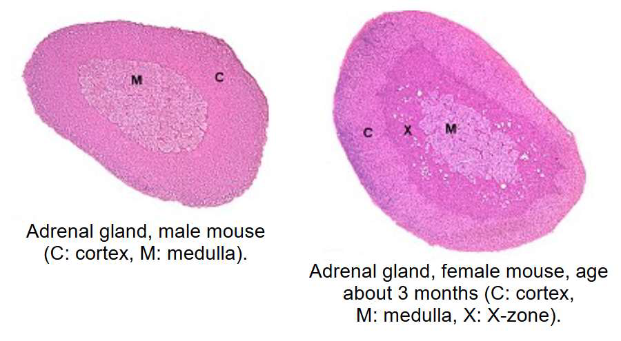
Adrenal gland
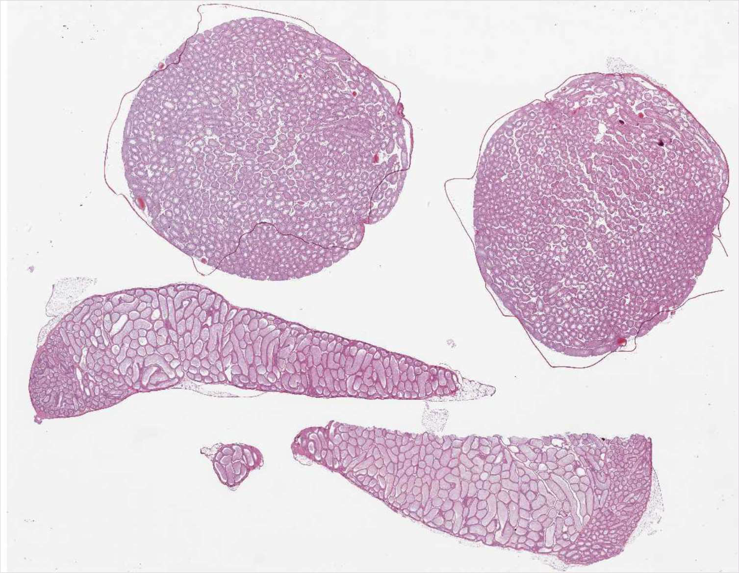
Testes
Testes and epididymides
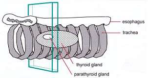
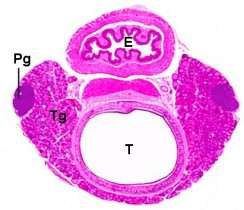
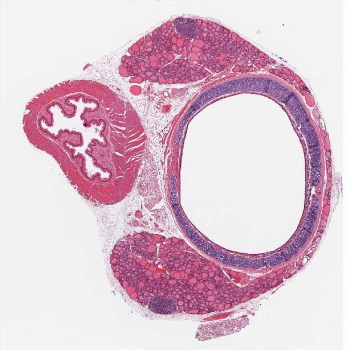
Thyroid gland
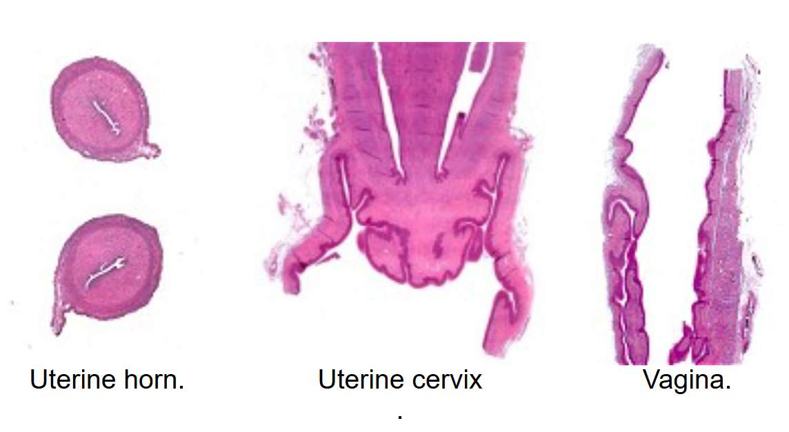
Uterus
Pathology
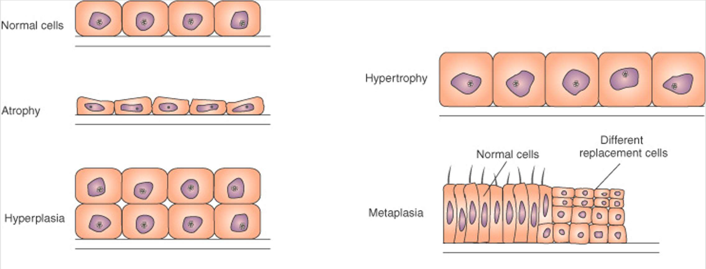
Cell increase
- Hypertrophy
size increase - Hyperplasia
number increase - Neoplasia
- Benign
- Malignant
- Hypertrophy

Cell death
- Necrosis
- “Unprogrammed” cell death
- Caused by external factors (infection, trauma, toxicity)
- Always detrimental
- Apoptosis
- Programmed cell death
- Initiated by internal signals
- Often healthy process, usually beneficial
- Necrosis
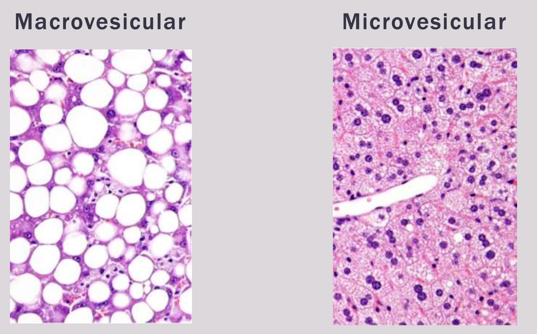
Vacuolation
- Glycogen
- Lipids
- Degeneration (hydropic change)
- Physiological
Liver pathology
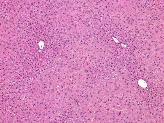
Enzyme induction
Fatty change
Necrosis
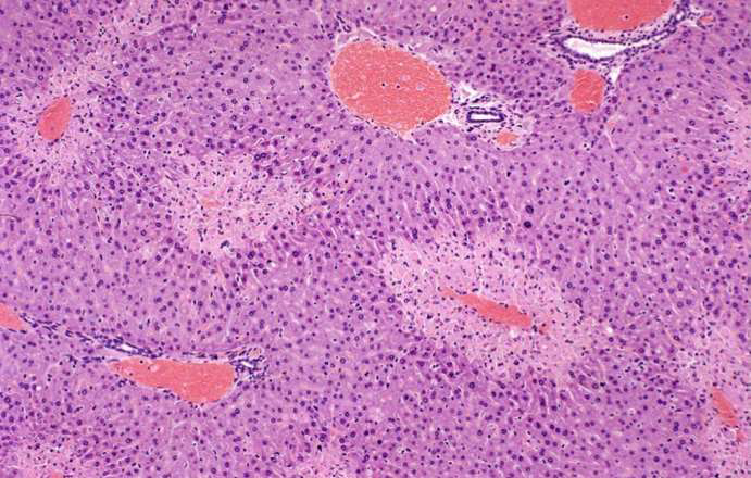
FOCAL NECROSIS
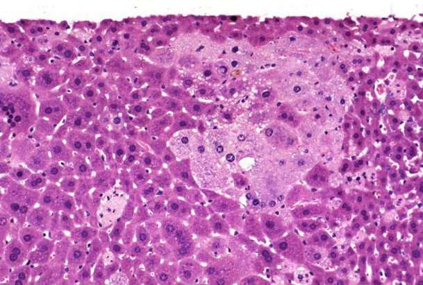
ZONAL NECROSIS
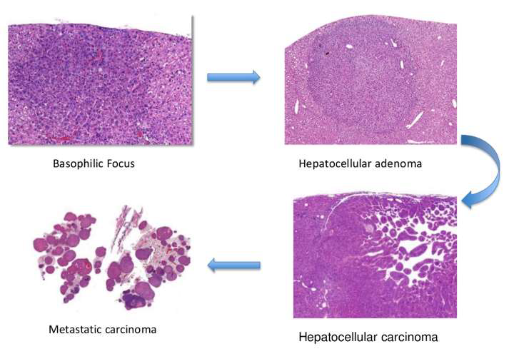
Tumour
Previous exam 2023
Organ Identification
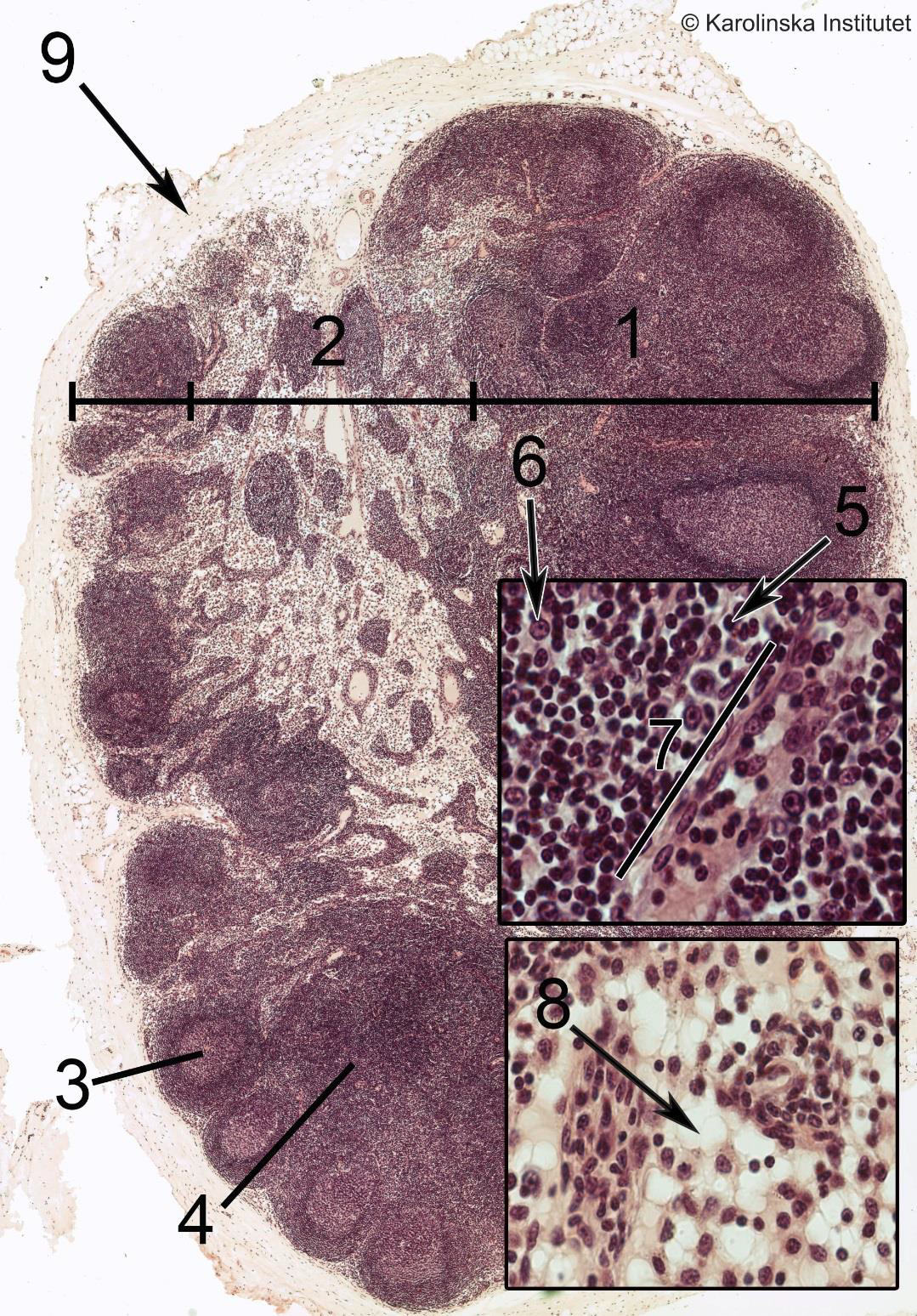
Lymph node
1
2
3
4
5
6
1. Cortex,
2. Medulla,
3. Germinal center,
4. Cortex,
5. Lymphocyte,
9. CapsulaStomach
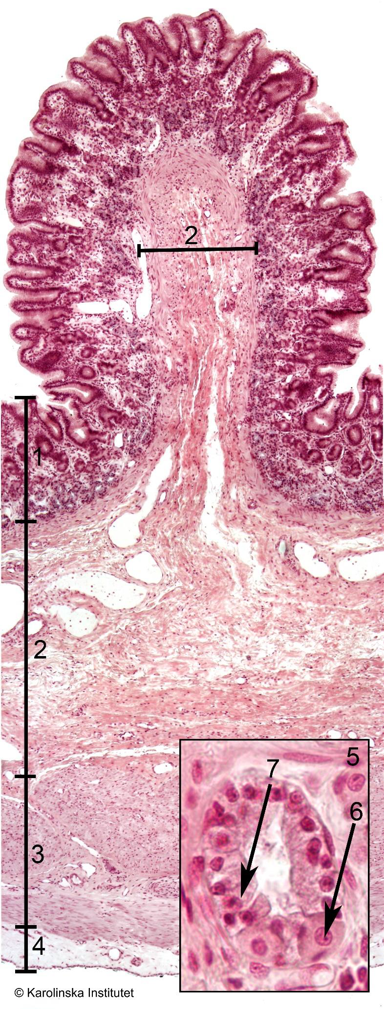
1
2
3
4
5
1. Mucosa,
2. Submucosa,
3. Muscularis,
4. Fibrosa,
6. Parietal cell,Stomach
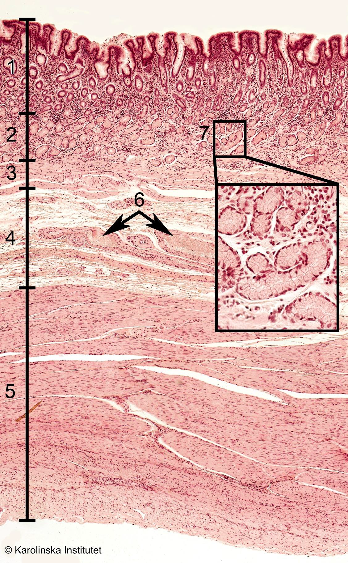
Duodenum
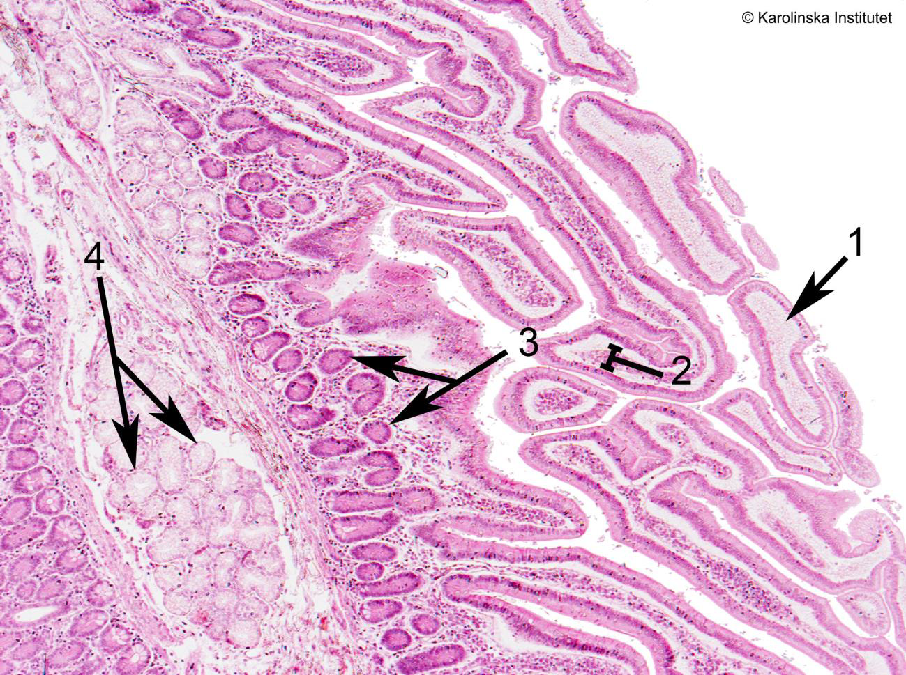
1
2
3
4
1. Villi (cross-cut),
2. Lamina propria,
3. Glands,
4. GlandsIleum
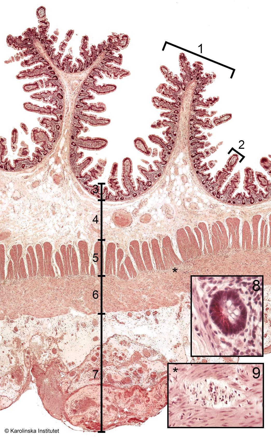
1
2
3
4
5
6
7
2. Villi,
3. Mucosa,
4. Submucosa,
5. Muscularis,
6. Muscularis,
7. Serosa,
8. Paneth cellsColon
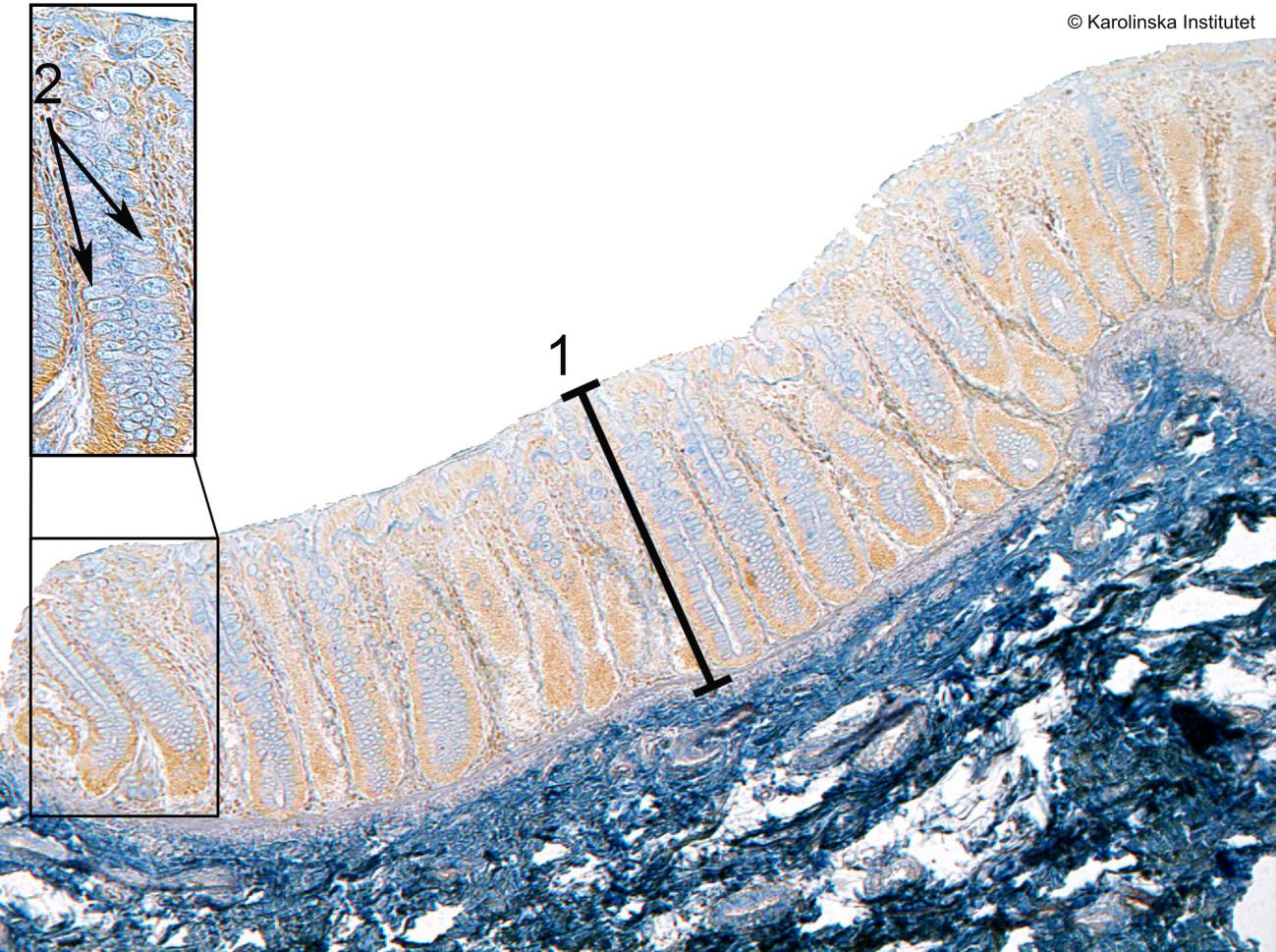
1
2
1. Crypt,
2. Goblet cellsLiver
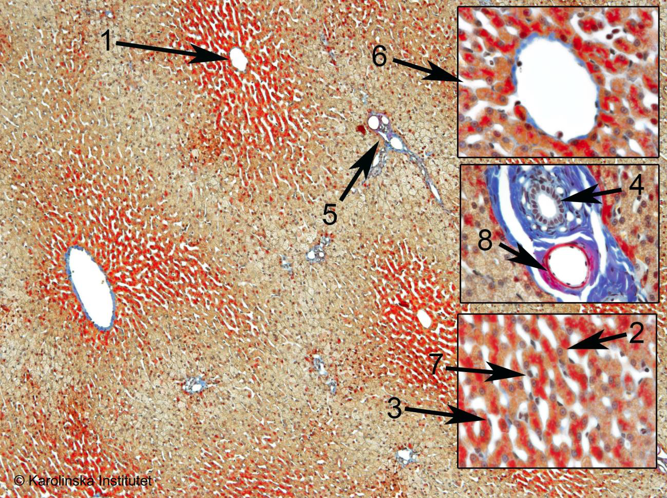
1
2
3
4
5
6
7
1. Central vein,
2. Hepatocyte,
3. Endothelial cell,
4. Bile duct,
5. Portal triad,
6. Sinusoid,
8. Blood vessel (in portal triad)Thyroid gland
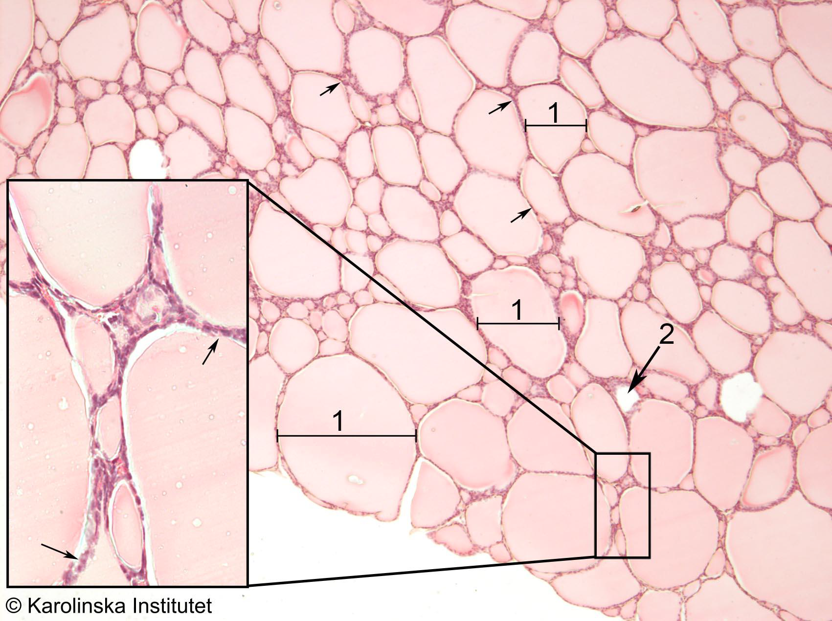
1
2
1. Follicle,
2. Blood vesselAdrenal gland
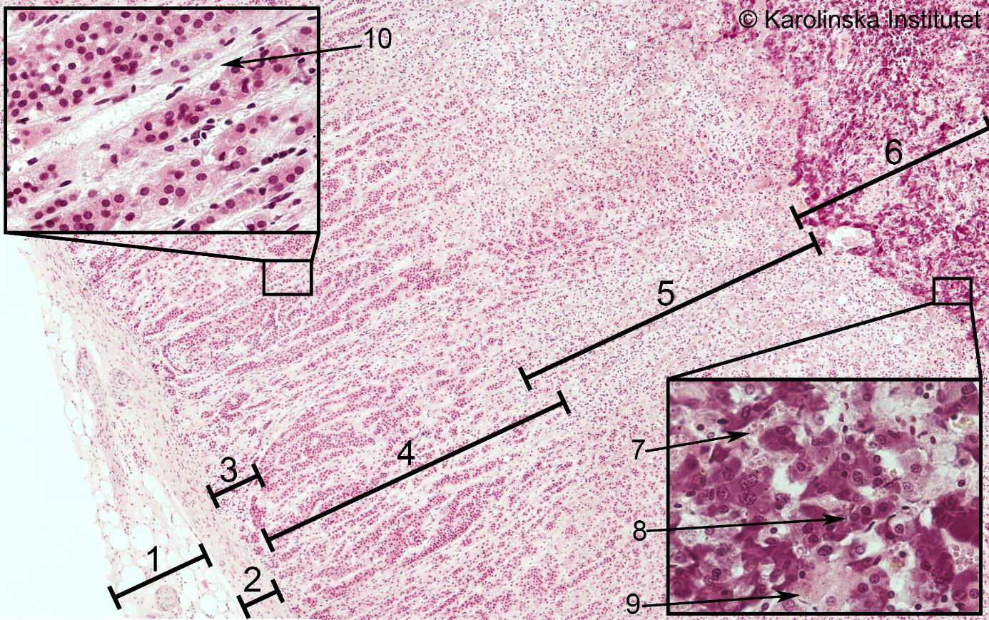
1
2
3
4
3. Zona
4. Zona
5. Zona
6. MedullaLung
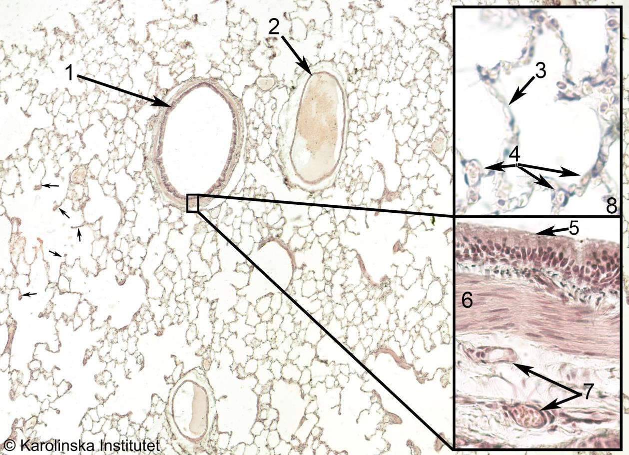
1
2
3
4
5
6
7
1. Bronchiole,
2. Vein,
3. Alveolar wall,
4. Capillary,
6. Smooth muscle,
7. Blood vessel,
8. AlveoliKidney
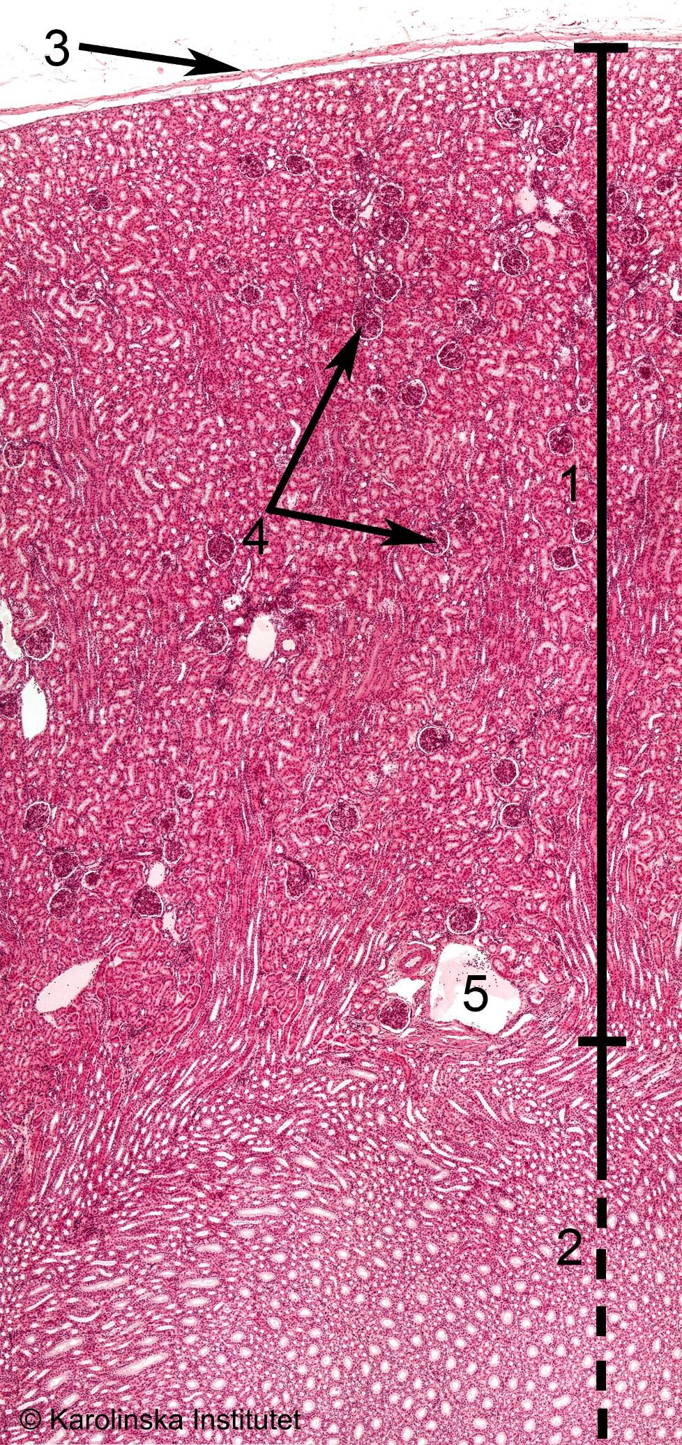
1
2
3
4
1. Cortex,
2. Medulla,
3. Capsula,
4. Glomeruli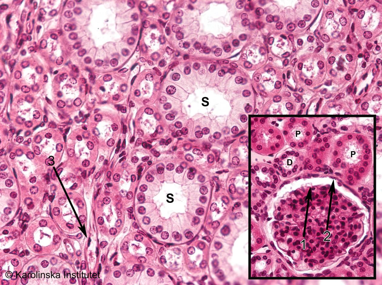
1
2. Bowman’s capsule
Testis
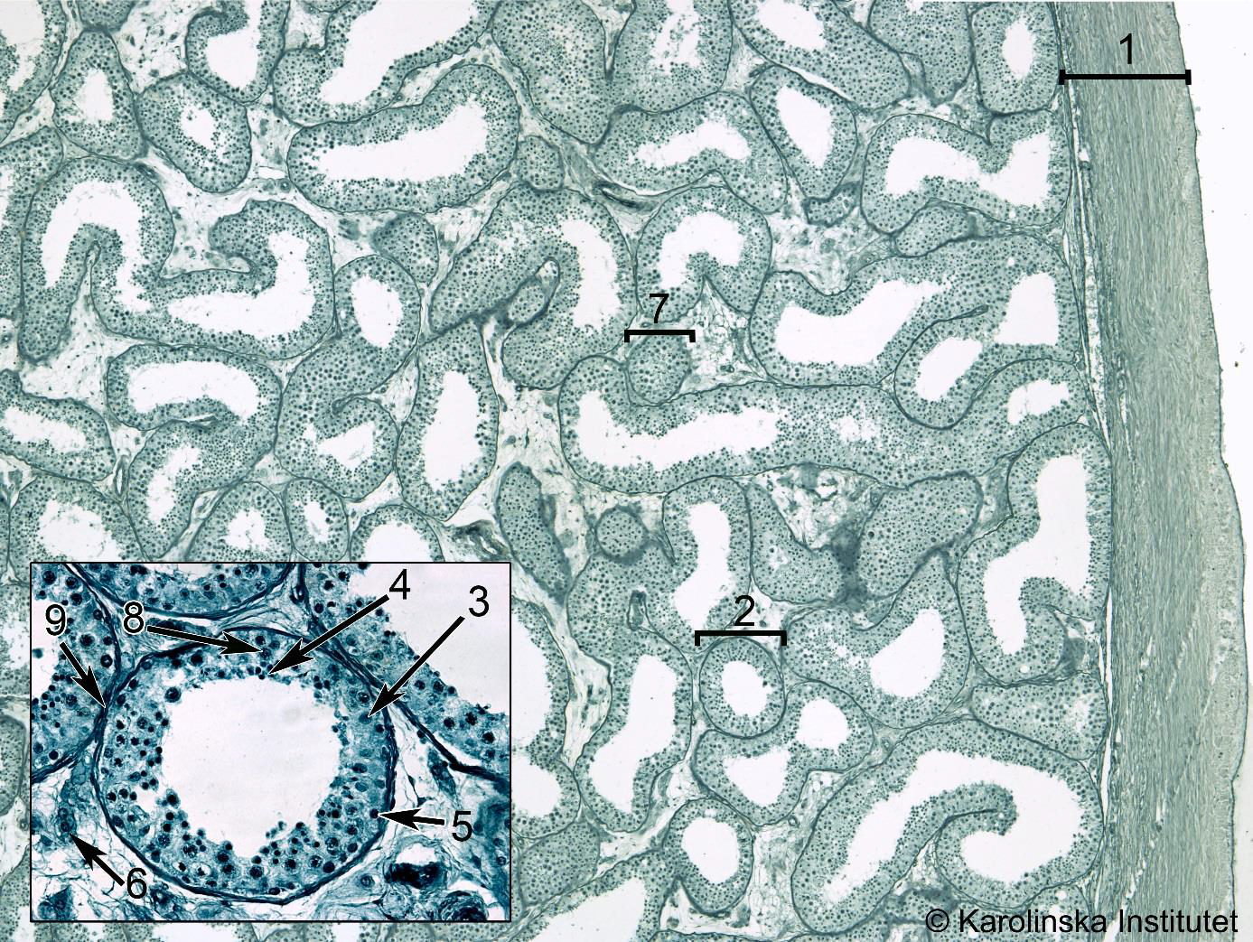
1
2
3
4
5
6
1. Tunica/capsule,
2. Tubuli,
3. Sertoli cell,
4. Spermatide,
5. Spermatogonium,
6. Leydig cellUterus
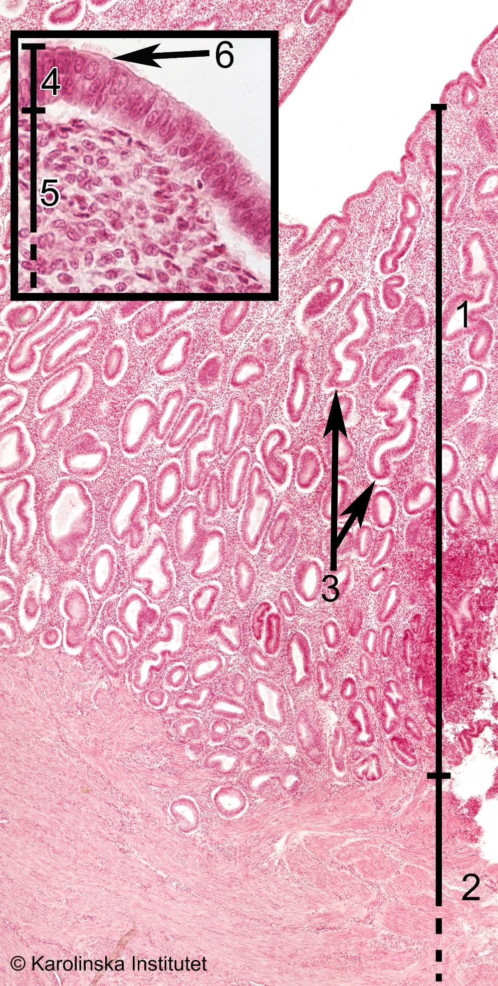
1
2
3
4
1. Endometrium,
2. Myometrium,
3. Glands,
4. EpitheliumQuestions
What is hemoglobin, in which cells can you find it and what is the physiological function of it? (3)
1
Hemoglobin is a protein in red blood cells responsible for transporting oxygen from the lungs to tissues and carrying carbon dioxide back to the lungs. It also helps regulate blood pH.
From ChatGPT
What is reticulocytes? How can you identify them? What could be a reason
behind an increased number of reticulocytes? (3)
1
2
3
4
5
6
7
Reticulocytes are immature red blood cells recently released from the bone marrow into the bloodstream. They still contain residual RNA.
Identification:
Identified using supravital stains (e.g., new methylene blue), which highlight their RNA as a reticular network.
Increased Numbers (Reticulocytosis):
Caused by increased erythropoiesis, commonly due to anemia (e.g., hemolytic anemia or acute blood loss) or recovery from bone marrow suppression.From ChatGPT
The following biomarkers could indicate liver damage: ALT, AST, ALP and total bilirubin. (4)
a. Which of these biomarkers is most specific for damaged hepatocytes? (2)
b. Which part of the liver is damaged if you find increased levels of ALP and total bilirubin? (2)1
2
3a. Alanine aminotransferase (ALT) is the most specific biomarker for damaged hepatocytes.
b. Increased levels of alkaline phosphatase (ALP) and total bilirubin indicate damage to the bile ducts or cholestasis, often associated with biliary obstruction.From ChatGPT and proved by slides and https://www.hepatitis.va.gov/HEPATITIS/course/index.asp?page=/provider/courses/livertests/livertests-10&
There is a lack of specific biomarkers for some organs. Mention one of these organs and explain briefly why you think it is difficult to measure toxicity in this organ from blood samples. (2)
1
The **respiratory tract** lacks specific biomarkers because it is exposed to both internal and external environments, leading to non-specific inflammatory responses. Many biomarkers associated with respiratory damage, like inflammatory cytokines or enzymes, are also found in systemic conditions, making it difficult to attribute changes specifically to respiratory toxicity from blood samples.
Explained by ChatGPT based on point from slides
Why is the time of day for blood sampling important in clinical pathology? (1)
1
The time of day for blood sampling is important because circadian rhythms affect levels of hormones, metabolites, and blood cells. Standardizing sampling times ensures consistent and accurate results.
From ChatGPT
In cases of major inflammation you can sometimes see decreased levels of albumin in the blood. What could be a reason behind that decrease? (2)
1
2
3
4In cases of major inflammation, decreased albumin levels can occur due to:
1. Acute-phase response: The liver prioritizes producing other proteins like fibrinogen and C-reactive protein, reducing albumin synthesis.
2. Increased capillary permeability: Inflammation can lead to leakage of albumin into the tissues, lowering blood levels.From ChatGPT
Artifacts. (3)
a. What is meant by “artifact”? (1)
b. Briefly describe one example of an artifact that can occur in tissue slides for light microscopy. (2)1
2
3a. An artifact in slide preparation refers to any distortion or alteration in the tissue sample that occurs during the process of fixation, embedding, sectioning, or staining, rather than being a true feature of the tissue.
b. An example of an artifact is air bubbles in tissue sections. These can form during the mounting process, causing gaps or distortions in the tissue, which may interfere with the clarity and interpretation of the slide under light microscopy.The following terms are common pathological terms. Briefly describe what they mean (compared to normal cells). (3)
a. Hypertrophy
b. Hyperplasia
c. Neoplasia1
2
3
4
5a. Hypertrophy: An increase in the size of individual cells, leading to enlargement of the tissue or organ, compared to normal cells, which remain a standard size.
b. Hyperplasia: An increase in the number of cells within a tissue or organ, resulting in tissue enlargement, compared to normal, steady cell proliferation.
c. Neoplasia: Uncontrolled, abnormal cell growth that forms a mass or tumor, unlike normal cells that grow and divide in a regulated manner.Tumours can be either benign or malignant. (4)
a. Which form is the most severe? (1)
b. Mention 3 characteristics of a benign tumour (as opposed to a malignant tumour). (3)1
2
3
4
5
6a. Malignant tumors are the most severe due to their potential to invade surrounding tissues and metastasize to other parts of the body.
b. Three characteristics of a benign tumor (as opposed to malignant):
1. Slow growth.
2. Well-defined borders, typically not invasive.
3. Does not metastasize to other parts of the body.During necropsy gross pathology findings can sometimes be observed for example in the liver. Give one example of such a finding, how it is described during necropsy and what could be the cause of this finding (what you may find when you look in the microscope). (3)
1
2
3
4
5
6
7Example: Fatty liver (Hepatic steatosis)
- Description during necropsy: The liver appears enlarged, pale, and has a greasy or yellowish appearance due to the accumulation of fat.
- Cause: Fatty liver can result from various causes such as alcohol abuse, obesity, or diabetes.
- Microscopic finding: The liver cells show intracellular fat droplets (mainly triglycerides) within the hepatocytes, which can displace the nucleus to the periphery.From ChatGPT
What does “plane of section” mean? Why is that important in histology? Give one example. (3)
1
2
3
4
5
6
7
8The plane of section refers to the direction in which a tissue sample is cut for examination. It's important in histology because it determines how the tissue structures are visualized.
Example:
- A longitudinal section cuts along the length of the tissue, revealing the full structure (e.g., muscle fibers).
- A cross section cuts perpendicular to the length, showing a slice of the tissue at a specific point (e.g., a cross-section of a blood vessel).
- An oblique section cuts at an angle, providing a view that may reveal features not seen in other sections.
The choice of section affects how tissue features are interpreted.From ChatGPT
Unfamiliar words
1
2
3
4
5
6
7
8
9
10
11
12
13
14
15
16
17
18
19
20
21germinal center (in lymph node)
muscularis
fibrosa (in stomach)
parietal cell (in stomach)
Duodenum
villi
lamina propria
ileum
serosa (in ileum)
paneth cell (in ileum)
goblet cell (in colon)
sinusoid (in liver)
bronchiole (in lung)
capillary (in lung)
glomeruli (in kidney)
sertoli cell (in testes)
spermatide (in testes)
spermatogonium (in testes)
leydig cell (in testes)
esophagus (in thyroid gland)
trachea (in thyroid gland)